Encefalopatía Neonatal: Revisión del Diagnóstico y Tratamiento.
La encefalopatía neonatal es un síndrome heterogéneo, clínicamente definido, caracterizado por una alteración de la función neurológica en los primeros días de vida en un bebé nacido a las 35 semanas de gestación o después de este periodo, que se manifiesta por un nivel bajo de alerta o convulsiones, a menudo acompañado de dificultad para iniciar y mantener la respiración, y por depresión del tono muscular y reflejos.
La encefalopatía neonatal puede deberse a una amplia variedad de etiologías. La encefalopatía hipóxico-isquémica es responsable únicamente de algunos de los casos de encefalopatía neonatal. Dado que la naturaleza subyacente de la lesión cerebral que causa el deterioro neurológico en un recién nacido a menudo no se conoce bien, la “encefalopatía neonatal” se ha convertido en el término de elección para describir el síndrome clínico de disfunción del sistema nervioso central en el período neonatal porque no implica una etiología o fisiopatología específica de base.
Definir si un evento hipóxico-isquémico agudo contribuyó a la encefalopatía neonatal es todo un desafío, ya que no existe una prueba estándar de oro para el diagnóstico. Los diversos signos clínicos de encefalopatía hipóxico-isquémica, que incluyen puntuaciones bajas de Apgar, pH bajo del cordón umbilical, convulsiones neonatales y encefalopatía, son inespecíficos y pueden ocurrir en ausencia de lesión cerebral hipóxico-isquémica global o secuelas neurológicas a largo plazo.
Sin embargo, cuando los síntomas clínicos sugieren que la encefalopatía hipóxico-isquémica es la causa más probable de encefalopatía neonatal, un “diagnóstico presuntivo de encefalopatía hipóxico-isquémica” suele ser adecuado, mientras se esperan resultados de pruebas adicionales, y al mismo tiempo se establecen terapias neuroprotectoras diseñadas específicamente para tratar la encefalopatía hipóxico-isquémica.
Factores de Riesgo de la Encefalopatía Neonatal
La mayoría de los casos de encefalopatía neonatal se asocian con factores de riesgo que se presentan previo al trabajo de parto. Éstos incluyen:
- Factores maternos, incluido el desempleo, antecedentes familiares de convulsiones o trastornos neurológicos, tratamiento de infertilidad y enfermedad de la tiroides.
- Afecciones placentarias, que incluyen preeclampsia grave y apariencia anormal de la placenta
- Alteraciones fetales, como la restricción del crecimiento intrauterino.
De estos, la restricción del crecimiento intrauterino es el factor de riesgo más importante. Los factores de riesgo intraparto para la encefalopatía neonatal incluyen los siguientes:
- Eventos intraparto agudos o eventos centinela (p.e. ruptura uterina, desprendimiento de placenta normoinserta, prolapso del cordón umbilical, circular de cordón, shock materno/muerte).
- Procesos inflamatorios (p.e. fiebre materna, corioamnionitis, ruptura prematura de membranas prolongada).
- Persistencia de la posición occipitoposterior.
- Distocia de hombros.
- Cesárea de emergencia.
- Parto vaginal instrumentado.
Un evento intraparto agudo (es decir, centinela), como un desprendimiento de la placenta o una ruptura uterina, confiere un mayor riesgo, pero está presente en sólo una minoría de niños con encefalopatía neonatal.
Presentación Clínica de la Encefalopatía Neonatal
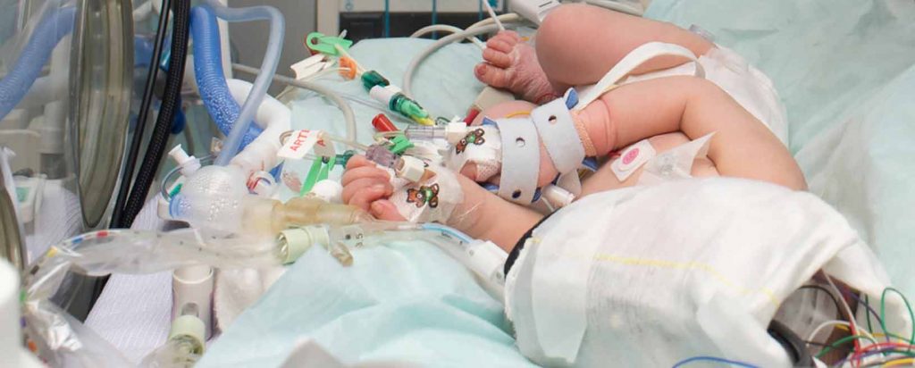
El neonato con encefalopatía se presenta con alteración del estado de alerta (por ejemplo, agitado, irritable, letárgico u obnubilado), con disminución de movimientos espontáneos, dificultad respiratoria o para la alimentación, tono muscular deficiente, postura anormal, ausencia de reflejos primitivos o convulsiones. En la sala de parto, el bebé a menudo exhibirá puntajes de Apgar bajos y un llanto débil o ausente. La gravedad de la encefalopatía neonatal se puede clasificar en leve, moderada o grave según estos hallazgos clínicos. Los recién nacidos con encefalopatía neonatal de moderada a grave a menudo requieren reanimación neonatal inmediata.
Diagnóstico de Encefalopatía neonatal
El diagnóstico de encefalopatía neonatal requiere la evaluación de las posibles etiologías. El Colegio Americano de Obstetras y Ginecólogos (ACOG) recomienda una evaluación integral en todos los casos de encefalopatía neonatal. Esta evaluación debe incluir el estado clínico neonatal y la consideración de todos los factores que potencialmente contribuyen a la encefalopatía neonatal, incluidos los antecedentes médicos maternos, antecedentes obstétricos, factores intraparto (incluidos los resultados de monitoreo de la frecuencia cardíaca fetal y los eventos centinela agudos) y la patología placentaria.
La presencia de oliguria, cardiomiopatía o pruebas de función hepática anormales pueden sugerir un evento hipóxico-isquémico global. La neuroimagen desempeña un papel clave en la evaluación de la encefalopatía neonatal y puede proporcionar información sobre la naturaleza, el patrón y la gravedad de la lesión cerebral. Se recomienda realizar una historia clínica materna y familiar completa, que incluya antecedentes de trastornos tromboembólicos, pérdida previa de embarazos, infección materna y uso de drogas por parte de la madre.
Los trastornos metabólicos, los olores inusuales, las características dismórficas y las anomalías congénitas pueden sugerir la presencia de un error innato del metabolismo o trastorno genético. La encefalopatía neonatal es probablemente secundaria a un evento hipóxico-isquémico agudo cuando una o más de las siguientes condiciones están presentes:
- Signos neonatales consistentes con un evento agudo hipóxico-isquémico periparto o intraparto:
- Puntuación de Apgar <5 a los 5 y 10 minutos
- pH de la arteria umbilical fetal <7.0, o déficit de base ≥ 12 mmol/L, o ambos
- Lesión cerebral aguda observada en RM cerebral o espectroscopia de resonancia magnética (MRS) compatible con evento hipóxico-isquémico.
- Presencia de insuficiencia orgánica multisistémica compatible con encefalopatía hipóxico-isquémica.
- Factores adicionales consistentes con un evento hipóxico-isquémico intraparto o periparto agudo:
- Un evento hipóxico o isquémico centinela inmediatamente antes o durante el trabajo de parto y/o el parto, como la ruptura uterina o desprendimiento grave de placenta.
- Patrones de monitorización de la frecuencia cardíaca fetal consistentes con un evento periparto o intraparto agudo, como un patrón de categoría III.
- La neuroimagen revela un patrón de lesión cerebral que es típico de lesión hipóxico-isquémica en el recién nacido, incluida la sustancia gris nuclear o lesión en la zona marginal.
Estudios que serán de utilidad
Se recomienda realizar las siguientes pruebas para evaluar la etiología de la encefalopatía neonatal según el criterio clínico y diagnóstico presuntivo. Se deben tomar muestras de sangre del cordón umbilical para determinar el pH arterial y venoso y el déficit de la base. Además, un examen macroscópico e histológico de la placenta y el cordón umbilical puede proporcionar evidencia de una causa contribuyente, como una lesión vascular placentaria o una infección, inflamación, o una trombosis del cordón umbilical. Solicita biometría hemática completa con diferencial para evaluar una posible infección, hemorragia y/o trombocitopenia.
Gasometría arterial, calcio, magnesio, glucosa y electrolitos en suero para guiar el manejo. Las enzimas hepáticas y la creatinina sérica pueden ayudar a identificar lesiones en otros órganos terminales. Los hemocultivos bacterianos para descartar sepsis y cultivos virales si existen datos sugerentes. Se debe realizar pruebas de coagulación, como el tiempo de protrombina (TP), el tiempo parcial de tromboplastina (TPT) y dímero D, si hay sangrado o supuración para descartar una coagulopatía intravascular diseminada.
Electroencefalografía
La electroencefalografía (EEG) ayudará a determinar si hay convulsiones clínicas o electrográficas y para evaluar la actividad eléctrica de fondo, ya que estos hallazgos pueden afectar el tratamiento y pronóstico de la encefalopatía neonatal. Generalmente, el EEG se obtiene en el primer día de vida (antes o durante el tratamiento) y la monitorización se continúa durante al menos 24 horas o más si hay convulsiones electrográficas. La EEG integrada de amplitud es una herramienta útil de monitorización continua que puede detectar la actividad convulsiva en bebés con encefalopatía neonatal y proporcionar información adicional sobre la actividad eléctrica de base.
Resonancia magnética cerebral
Se realiza entre los cuatro y siete días de edad. Los hallazgos específicos en la RM cerebral pueden ser útiles para determinar la patogenia y el pronóstico de la encefalopatía neonatal. La ecografía craneal no es tan sensible como la RM, y la TAC de cráneo en el período neonatal agudo tiene poca sensibilidad para la detección de lesiones e impone una exposición a la radiación evitable.
Pruebas metabólicas
Pruebas específicas para el error innato del metabolismo, incluido el amoníaco, el lactato y el piruvato, los aminoácidos séricos y los ácidos orgánicos en orina para descartar una causa metabólica de encefalopatía neonatal. Se sugieren pruebas genéticas (por ejemplo, citogenética y microarray de hibridación genómica comparativa) si el niño es dismórfico o presenta anomalías congénitas.
Punción lumbar
Si existe sospecha de infección intracraneal (p.e. fiebre, leucocitosis, exantema, hemocultivo positivo y/o lesión de herpes materna o infección documentada), ya que la meningitis puede simular los signos y síntomas de la encefalopatía neonatal. Los antibióticos se inician hasta que se confirma la infección, y se inicia el aciclovir si se sospecha del virus del herpes simple.
Tratamiento de la Encefalopatía Neonatal
La hipotermia terapéutica es el tratamiento de elección (en las primeras seis horas de vida) para la encefalopatía neonatal que cumple con los criterios para la presunta encefalopatía hipóxico-isquémica. Las medidas de soporte de la encefalopatía neonatal moderada y grave debe realizarse en una unidad de cuidados intensivos neonatales. Los objetivos principales incluyen el mantenimiento de la homeostasis fisiológica y el tratamiento de las manifestaciones externas de la lesión cerebral. Los aspectos centrales de las medidas de soporte incluyen los siguientes:
- Mantenimiento de una ventilación adecuada (evitar hipoxia o hiperoxia).
- Mantenimiento de perfusión cerebral y de órganos adecuada (evitar la hipotensión sistémica o la hipertensión; evitar la hiperviscosidad).
- Mantenimiento de un estado metabólico normal (p.e. normoglucemia, estado nutricional, pH).
- Prevención y/o control de las convulsiones.
- Prevención y/o control del edema cerebral (evitar la sobrecarga de líquidos)
Hipotermia Terapéutica
La hipotermia terapéutica, mantenida durante 72 horas a una temperatura de 33 a 35°C e iniciada dentro de las primeras seis horas después del parto, es la única terapia neuroprotectora comprobada para el tratamiento de la encefalopatía neonatal. Existe un consenso entre expertos de que la hipotermia terapéutica debería estar disponible de manera universal, en congruencia con el beneficio y la seguridad de la hipotermia, y la falta de otros tratamientos efectivos. Por ello, la hipotermia se ha convertido en el estándar de tratamiento en la mayoría de las unidades de cuidados intensivos neonatales en los Estados Unidos, Europa, Australia y Japón. En general, los criterios de elegibilidad para la hipotermia terapéutica incluyen los siguientes:
- Edad gestacional ≥36 semanas y ≤6 horas de edad.
- Un pH ≤7.0 o un déficit de base de ≥16 mmol/L en una muestra de sangre del cordón umbilical o cualquier sitio obtenida en la primera hora después del nacimiento
- Uno de los siguientes:
- Una puntuación de Apgar <5 a los 10 minutos.
- Reanimación neonatal contínua (p.e. ventilación asistida, compresiones torácicas o vasopresores) iniciada al nacer y continuada durante al menos 10 minutos.
- Encefalopatía moderada a grave en el examen clínico.
Se ha demostrado que la hipotermia terapéutica únicamente mejora los resultados en lactantes con encefalopatía moderada a grave. Se desconoce si este tratamiento mejora los resultados en lactantes con grados más leves de encefalopatía. La mayoría de los centros utilizan la escala de Sarnat modificada, con o sin información adicional sobre la presencia de convulsiones. La prueba de Sarnat evalúa la gravedad de las alteraciones del nivel de alerta, la actividad espontánea, el tono, la postura, los reflejos primitivos y la función autónoma.
Medidas de Soporte
Además del tratamiento con hipotermia terapéutica, el tratamiento sugerido para la encefalopatía neonatal incluye las siguientes recomendaciones:
- Tratamiento de las convulsiones con fenobarbital, lorazepam, fosfenitoína o levetiracetam. El agente terapéutico óptimo, así como la duración del tratamiento, no se ha evaluado adecuadamente.
- Ventilación de alta frecuencia, el óxido nítrico o las terapias de oxigenación con membrana extracorpórea, según estén disponibles, para bebés con hipertensión pulmonar persistente para mantener la oxigenación.
- Sustitución de volumen y uso de agentes inotrópicos según sea necesario para mantener la presión arterial y una perfusión cerebral adecuada. Sin embargo, debe evitarse la hipertensión sistémica y la sobrecarga de volumen, que pueden empeorar el edema cerebral.
- Tratamiento precoz del trastorno metabólico. Debe interrumpirse la alimentación, corregir la acidosis y la hipoglucemia, y considerar un tratamiento específico como la suplementación o la hemodiálisis después de consultar con un genetista o un especialista en metabolismo pediátrico.
Cuando no se utiliza la hipotermia terapéutica, se recomienda una estrecha vigilancia de la temperatura corporal central y evitar la hipertermia, ya que está relacionada con resultados adversos para el paciente.
Referencias Bibliográficas
Executive summary: Neonatal encephalopathy and neurologic outcome, second edition. Report of the American College of Obstetricians and Gynecologists’ Task Force on Neonatal Encephalopathy. Obstet Gynecol 2014; 123:896.
Volpe JJ. Neonatal encephalopathy: an inadequate term for hypoxic-ischemic encephalopathy. Ann Neurol 2012; 72:156.
Wu YW, Backstrand KH, Zhao S, et al. Declining diagnosis of birth asphyxia in California: 1991-2000. Pediatrics 2004; 114:1584.
Graham EM, Ruis KA, Hartman AL, et al. A systematic review of the role of intrapartum hypoxia-ischemia in the causation of neonatal encephalopathy. Am J Obstet Gynecol 2008; 199:587.
Thornberg E, Thiringer K, Odeback A, Milsom I. Birth asphyxia: incidence, clinical course and outcome in a Swedish population. Acta Paediatr 1995; 84:927.
Lee AC, Kozuki N, Blencowe H, et al. Intrapartum-related neonatal encephalopathy incidence and impairment at regional and global levels for 2010 with trends from 1990. Pediatr Res 2013; 74 Suppl 1:50.
Kurinczuk JJ, White-Koning M, Badawi N. Epidemiology of neonatal encephalopathy and hypoxic-ischaemic encephalopathy. Early Hum Dev 2010; 86:329.
Chau V, Poskitt KJ, Miller SP. Advanced neuroimaging techniques for the term newborn with encephalopathy. Pediatr Neurol 2009; 40:181.
Barnette AR, Horbar JD, Soll RF, et al. Neuroimaging in the evaluation of neonatal encephalopathy. Pediatrics 2014; 133:e1508.
Redline RW. Severe fetal placental vascular lesions in term infants with neurologic impairment. Am J Obstet Gynecol 2005; 192:452.
Miller SP, Ramaswamy V, Michelson D, et al. Patterns of brain injury in term neonatal encephalopathy. J Pediatr 2005; 146:453.
Miller SP, Newton N, Ferriero DM, et al. Predictors of 30-month outcome after perinatal depression: role of proton MRS and socioeconomic factors. Pediatr Res 2002; 52:71.
Barnett A, Mercuri E, Rutherford M, et al. Neurological and perceptual-motor outcome at 5 – 6 years of age in children with neonatal encephalopathy: relationship with neonatal brain MRI. Neuropediatrics 2002; 33:242.
Heinz ER, Provenzale JM. Imaging findings in neonatal hypoxia: a practical review. AJR Am J Roentgenol 2009; 192:41.
Ghei SK, Zan E, Nathan JE, et al. MR imaging of hypoxic-ischemic injury in term neonates: pearls and pitfalls. Radiographics 2014; 34:1047.
Ferriero DM. Neonatal brain injury. N Engl J Med 2004; 351:1985.
Barkovich AJ. MR and CT evaluation of profound neonatal and infantile asphyxia. AJNR Am J Neuroradiol 1992; 13:959.
Roland EH, Poskitt K, Rodriguez E, et al. Perinatal hypoxic-ischemic thalamic injury: clinical features and neuroimaging. Ann Neurol 1998; 44:161.
Okereafor A, Allsop J, Counsell SJ, et al. Patterns of brain injury in neonates exposed to perinatal sentinel events. Pediatrics 2008; 121:906.
Myers RE. Two patterns of perinatal brain damage and their conditions of occurrence. Am J Obstet Gynecol 1972; 112:246.
Gano D, Sargent MA, Miller SP, et al. MRI findings in infants with infantile spasms after neonatal hypoxic-ischemic encephalopathy. Pediatr Neurol 2013; 49:401.
Martinez-Biarge M, Bregant T, Wusthoff CJ, et al. White matter and cortical injury in hypoxic-ischemic encephalopathy: antecedent factors and 2-year outcome. J Pediatr 2012; 161:799.
Thayyil S, Chandrasekaran M, Taylor A, et al. Cerebral magnetic resonance biomarkers in neonatal encephalopathy: a meta-analysis. Pediatrics 2010; 125:e382.
Shankaran S, Kottamasu SR, Kuhns L. Brain sonography, computed tomography, and single-photon emission computed tomography in term neonates with perinatal asphyxia. Clin Perinatol 1993; 20:379.
Miller SP, Cozzio CC, Goldstein RB, et al. Comparing the diagnosis of white matter injury in premature newborns with serial MR imaging and transfontanel ultrasonography findings. AJNR Am J Neuroradiol 2003; 24:1661.
Gluckman PD, Wyatt JS, Azzopardi D, et al. Selective head cooling with mild systemic hypothermia after neonatal encephalopathy: multicentre randomised trial. Lancet 2005; 365:663.
Volpe JJ. Hypoxic-ischemic encephalopathy: clinical aspects. In: Neurology of the Newborn, 5th, Volpe JJ (Ed), Saunders, Philadelphia 2008. p.400.
Yager JY, Armstrong EA, Black AM. Treatment of the term newborn with brain injury: simplicity as the mother of invention. Pediatr Neurol 2009; 40:237.
Committee on Fetus and Newborn, Papile LA, Baley JE, et al. Hypothermia and neonatal encephalopathy. Pediatrics 2014; 133:1146.
Perlman M, Shah P. Time to adopt cooling for neonatal hypoxic-ischemic encephalopathy: response to a previous commentary. Pediatrics 2008; 121:616.
Azzopardi D, Strohm B, Edwards AD, et al. Treatment of asphyxiated newborns with moderate hypothermia in routine clinical practice: how cooling is managed in the UK outside a clinical trial. Arch Dis Child Fetal Neonatal Ed 2009; 94:F260.
Higgins RD, Raju T, Edwards AD, et al. Hypothermia and other treatment options for neonatal encephalopathy: an executive summary of the Eunice Kennedy Shriver NICHD workshop. J Pediatr 2011; 159:851.
National Institute for Health and Clinical Excellence. IPG347. Therapeutic hypothermia with intracorporeal temperature monitoring for hypoxic perinatal brain injury.
Takenouchi T, Iwata O, Nabetani M, Tamura M. Therapeutic hypothermia for neonatal encephalopathy: JSPNM & MHLW Japan Working Group Practice Guidelines Consensus Statement from the Working Group on Therapeutic Hypothermia for Neonatal Encephalopathy, Ministry of Health, Labor and Welfare (MHLW), Japan, and Japan Society for Perinatal and Neonatal Medicine (JSPNM). Brain Dev 2012; 34:165.
Wyckoff MH, Aziz K, Escobedo MB, et al. Part 13: Neonatal Resuscitation: 2015 American Heart Association Guidelines Update for Cardiopulmonary Resuscitation and Emergency Cardiovascular Care. Circulation 2015; 132:S543.
Perlman JM, Wyllie J, Kattwinkel J, et al. Part 7: Neonatal Resuscitation: 2015 International Consensus on Cardiopulmonary Resuscitation and Emergency Cardiovascular Care Science With Treatment Recommendations. Circulation 2015; 132:S204.
Sarnat HB, Sarnat MS. Neonatal encephalopathy following fetal distress. A clinical and electroencephalographic study. Arch Neurol 1976; 33:696.
Tagin MA, Woolcott CG, Vincer MJ, et al. Hypothermia for neonatal hypoxic ischemic encephalopathy: an updated systematic review and meta-analysis. Arch Pediatr Adolesc Med 2012; 166:558.
Jacobs SE, Berg M, Hunt R, et al. Cooling for newborns with hypoxic ischaemic encephalopathy. Cochrane Database Syst Rev 2013; :CD003311.
Shankaran S, Pappas A, McDonald SA, et al. Childhood outcomes after hypothermia for neonatal encephalopathy. N Engl J Med 2012; 366:2085.
Azzopardi D, Strohm B, Marlow N, et al. Effects of hypothermia for perinatal asphyxia on childhood outcomes. N Engl J Med 2014; 371:140.
Papile LA. Systemic hypothermia–a “cool” therapy for neonatal hypoxic-ischemic encephalopathy. N Engl J Med 2005; 353:1619.
Higgins RD, Raju TN, Perlman J, et al. Hypothermia and perinatal asphyxia: executive summary of the National Institute of Child Health and Human Development workshop. J Pediatr 2006; 148:170.
Blackmon LR, Stark AR, American Academy of Pediatrics Committee on Fetus and Newborn. Hypothermia: a neuroprotective therapy for neonatal hypoxic-ischemic encephalopathy. Pediatrics 2006; 117:942.
Laptook AR, Shankaran S, Tyson JE, et al. Effect of Therapeutic Hypothermia Initiated After 6 Hours of Age on Death or Disability Among Newborns With Hypoxic-Ischemic Encephalopathy: A Randomized Clinical Trial. JAMA 2017; 318:1550.
Akula VP, Joe P, Thusu K, et al. A randomized clinical trial of therapeutic hypothermia mode during transport for neonatal encephalopathy. J Pediatr 2015; 166:856.
Szakmar E, Kovacs K, Meder U, et al. Feasibility and Safety of Controlled Active Hypothermia Treatment During Transport in Neonates With Hypoxic-Ischemic Encephalopathy. Pediatr Crit Care Med 2017; 18:1159.
Shankaran S, Laptook AR, Ehrenkranz RA, et al. Whole-body hypothermia for neonates with hypoxic-ischemic encephalopathy. N Engl J Med 2005; 353:1574.
Azzopardi DV, Strohm B, Edwards AD, et al. Moderate hypothermia to treat perinatal asphyxial encephalopathy. N Engl J Med 2009; 361:1349.
Simbruner G, Mittal RA, Rohlmann F, et al. Systemic hypothermia after neonatal encephalopathy: outcomes of neo.nEURO.network RCT. Pediatrics 2010; 126:e771.
Jacobs SE, Morley CJ, Inder TE, et al. Whole-body hypothermia for term and near-term newborns with hypoxic-ischemic encephalopathy: a randomized controlled trial. Arch Pediatr Adolesc Med 2011; 165:692.
Zhou WH, Cheng GQ, Shao XM, et al. Selective head cooling with mild systemic hypothermia after neonatal hypoxic-ischemic encephalopathy: a multicenter randomized controlled trial in China. J Pediatr 2010; 157:367.
Gunn AJ, Gluckman PD, Gunn TR. Selective head cooling in newborn infants after perinatal asphyxia: a safety study. Pediatrics 1998; 102:885.
Shankaran S, Laptook AR, Pappas A, et al. Effect of depth and duration of cooling on deaths in the NICU among neonates with hypoxic ischemic encephalopathy: a randomized clinical trial. JAMA 2014; 312:2629.
Shankaran S, Laptook AR, Pappas A, et al. Effect of Depth and Duration of Cooling on Death or Disability at Age 18 Months Among Neonates With Hypoxic-Ischemic Encephalopathy: A Randomized Clinical Trial. JAMA 2017; 318:57.
Rossouw G, Irlam J, Horn AR. Therapeutic hypothermia for hypoxic ischaemic encephalopathy using low-technology methods: a systematic review and meta-analysis. Acta Paediatr 2015; 104:1217.
Woods AG, Cederholm CK. Subcutaneous fat necrosis and whole-body cooling therapy for neonatal encephalopathy. Adv Neonatal Care 2012; 12:345.
Wyatt JS, Gluckman PD, Liu PY, et al. Determinants of outcomes after head cooling for neonatal encephalopathy. Pediatrics 2007; 119:912.
Laptook A, Tyson J, Shankaran S, et al. Elevated temperature after hypoxic-ischemic encephalopathy: risk factor for adverse outcomes. Pediatrics 2008; 122:491.
Laptook AR, McDonald SA, Shankaran S, et al. Elevated temperature and 6- to 7-year outcome of neonatal encephalopathy. Ann Neurol 2013; 73:520.
Wu Q, Chen W, Sinha B, et al. Neuroprotective agents for neonatal hypoxic-ischemic brain injury. Drug Discov Today 2015; 20:1372.
Dixon BJ, Reis C, Ho WM, et al. Neuroprotective Strategies after Neonatal Hypoxic Ischemic Encephalopathy. Int J Mol Sci 2015; 16:22368.
Rangarajan V, Juul SE. Erythropoietin: emerging role of erythropoietin in neonatal neuroprotection. Pediatr Neurol 2014; 51:481.
Zhu C, Kang W, Xu F, et al. Erythropoietin improved neurologic outcomes in newborns with hypoxic-ischemic encephalopathy. Pediatrics 2009; 124:e218.
Wu YW, Mathur AM, Chang T, et al. High-Dose Erythropoietin and Hypothermia for Hypoxic-Ischemic Encephalopathy: A Phase II Trial. Pediatrics 2016; 137.
Malla RR, Asimi R, Teli MA, et al. Erythropoietin monotherapy in perinatal asphyxia with moderate to severe encephalopathy: a randomized placebo-controlled trial. J Perinatol 2017; 37:596.
Garg B, Sharma D, Bansal A. Systematic review seeking erythropoietin role for neuroprotection in neonates with hypoxic ischemic encephalopathy: presently where do we stand. J Matern Fetal Neonatal Med 2018; 31:3214.
Robertson C, Finer N. Term infants with hypoxic-ischemic encephalopathy: outcome at 3.5 years. Dev Med Child Neurol 1985; 27:473.
Robertson CM, Finer NN, Grace MG. School performance of survivors of neonatal encephalopathy associated with birth asphyxia at term. J Pediatr 1989; 114:753.
Finer NN, Robertson CM, Richards RT, et al. Hypoxic-ischemic encephalopathy in term neonates: perinatal factors and outcome. J Pediatr 1981; 98:112.
Levene ML, Kornberg J, Williams TH. The incidence and severity of post-asphyxial encephalopathy in full-term infants. Early Hum Dev 1985; 11:21.
van Handel M, Swaab H, de Vries LS, Jongmans MJ. Long-term cognitive and behavioral consequences of neonatal encephalopathy following perinatal asphyxia: a review. Eur J Pediatr 2007; 166:645.
Pappas A, Shankaran S, McDonald SA, et al. Cognitive outcomes after neonatal encephalopathy. Pediatrics 2015; 135:e624.
Martinello K, Hart AR, Yap S, et al. Management and investigation of neonatal encephalopathy: 2017 update. Arch Dis Child Fetal Neonatal Ed 2017; 102:F346.
Shankaran S, McDonald SA, Laptook AR, et al. Neonatal Magnetic Resonance Imaging Pattern of Brain Injury as a Biomarker of Childhood Outcomes following a Trial of Hypothermia for Neonatal Hypoxic-Ischemic Encephalopathy. J Pediatr 2015; 167:987.
Shankaran S, Woldt E, Koepke T, et al. Acute neonatal morbidity and long-term central nervous system sequelae of perinatal asphyxia in term infants. Early Hum Dev 1991; 25:135.
Lacey JL, Henderson-Smart DJ. Assessment of preterm infants in the intensive-care unit to predict cerebral palsy and motor outcome at 6 years. Dev Med Child Neurol 1998; 40:310.
Glass HC, Hong KJ, Rogers EE, et al. Risk factors for epilepsy in children with neonatal encephalopathy. Pediatr Res 2011; 70:535.
Kwon JM, Guillet R, Shankaran S, et al. Clinical seizures in neonatal hypoxic-ischemic encephalopathy have no independent impact on neurodevelopmental outcome: secondary analyses of data from the neonatal research network hypothermia trial. J Child Neurol 2011; 26:322.
Martinez-Biarge M, Diez-Sebastian J, Kapellou O, et al. Predicting motor outcome and death in term hypoxic-ischemic encephalopathy. Neurology 2011; 76:2055.
Alderliesten T, de Vries LS, Benders MJ, et al. MR imaging and outcome of term neonates with perinatal asphyxia: value of diffusion-weighted MR imaging and ¹H MR spectroscopy. Radiology 2011; 261:235.
van Laerhoven H, de Haan TR, Offringa M, et al. Prognostic tests in term neonates with hypoxic-ischemic encephalopathy: a systematic review. Pediatrics 2013; 131:88.
Massaro AN. MRI for neurodevelopmental prognostication in the high-risk term infant. Semin Perinatol 2015; 39:159.
Steinman KJ, Gorno-Tempini ML, Glidden DV, et al. Neonatal watershed brain injury on magnetic resonance imaging correlates with verbal IQ at 4 years. Pediatrics 2009; 123:1025.
Wu YW, Croen LA, Shah SJ, et al. Cerebral palsy in a term population: risk factors and neuroimaging findings. Pediatrics 2006; 118:690.
Barkovich AJ, Hajnal BL, Vigneron D, et al. Prediction of neuromotor outcome in perinatal asphyxia: evaluation of MR scoring systems. AJNR Am J Neuroradiol 1998; 19:143.
Massaro AN, Kadom N, Chang T, et al. Quantitative analysis of magnetic resonance images and neurological outcome in encephalopathic neonates treated with whole-body hypothermia. J Perinatol 2010; 30:596.
Barkovich AJ, Miller SP, Bartha A, et al. MR imaging, MR spectroscopy, and diffusion tensor imaging of sequential studies in neonates with encephalopathy. AJNR Am J Neuroradiol 2006; 27:533.
Rutherford M, Ramenghi LA, Edwards AD, et al. Assessment of brain tissue injury after moderate hypothermia in neonates with hypoxic-ischaemic encephalopathy: a nested substudy of a randomised controlled trial. Lancet Neurol 2010; 9:39.
Shankaran S, Barnes PD, Hintz SR, et al. Brain injury following trial of hypothermia for neonatal hypoxic-ischaemic encephalopathy. Arch Dis Child Fetal Neonatal Ed 2012; 97:F398.
Corbo ET, Bartnik-Olson BL, Machado S, et al. The effect of whole-body cooling on brain metabolism following perinatal hypoxic-ischemic injury. Pediatr Res 2012; 71:85.
Cheong JL, Coleman L, Hunt RW, et al. Prognostic utility of magnetic resonance imaging in neonatal hypoxic-ischemic encephalopathy: substudy of a randomized trial. Arch Pediatr Adolesc Med 2012; 166:634.
Inder TE, Hunt RW, Morley CJ, et al. Randomized trial of systemic hypothermia selectively protects the cortex on MRI in term hypoxic-ischemic encephalopathy. J Pediatr 2004; 145:835.
Azzopardi D, Edwards AD. Magnetic resonance biomarkers of neuroprotective effects in infants with hypoxic ischemic encephalopathy. Semin Fetal Neonatal Med 2010; 15:261.
Rutherford MA, Pennock JM, Counsell SJ, et al. Abnormal magnetic resonance signal in the internal capsule predicts poor neurodevelopmental outcome in infants with hypoxic-ischemic encephalopathy. Pediatrics 1998; 102:323.
de Vries LS, Jongmans MJ. Long-term outcome after neonatal hypoxic-ischaemic encephalopathy. Arch Dis Child Fetal Neonatal Ed 2010; 95:F220.
Perez A, Ritter S, Brotschi B, et al. Long-term neurodevelopmental outcome with hypoxic-ischemic encephalopathy. J Pediatr 2013; 163:454.
Harteman JC, Groenendaal F, Toet MC, et al. Diffusion-weighted imaging changes in cerebral watershed distribution following neonatal encephalopathy are not invariably associated with an adverse outcome. Dev Med Child Neurol 2013; 55:642.
Bonifacio SL, Saporta A, Glass HC, et al. Therapeutic hypothermia for neonatal encephalopathy results in improved microstructure and metabolism in the deep gray nuclei. AJNR Am J Neuroradiol 2012; 33:2050.
Robertson RL, Ben-Sira L, Barnes PD, et al. MR line-scan diffusion-weighted imaging of term neonates with perinatal brain ischemia. AJNR Am J Neuroradiol 1999; 20:1658.
Amess PN, Penrice J, Wylezinska M, et al. Early brain proton magnetic resonance spectroscopy and neonatal neurology related to neurodevelopmental outcome at 1 year in term infants after presumed hypoxic-ischaemic brain injury. Dev Med Child Neurol 1999; 41:436.
Hanrahan JD, Cox IJ, Azzopardi D, et al. Relation between proton magnetic resonance spectroscopy within 18 hours of birth asphyxia and neurodevelopment at 1 year of age. Dev Med Child Neurol 1999; 41:76.
Boichot C, Walker PM, Durand C, et al. Term neonate prognoses after perinatal asphyxia: contributions of MR imaging, MR spectroscopy, relaxation times, and apparent diffusion coefficients. Radiology 2006; 239:839.
Boardman JP, Ganesan V, Rutherford MA, et al. Magnetic resonance image correlates of hemiparesis after neonatal and childhood middle cerebral artery stroke. Pediatrics 2005; 115:321.
Mercuri E, Rutherford M, Cowan F, et al. Early prognostic indicators of outcome in infants with neonatal cerebral infarction: a clinical, electroencephalogram, and magnetic resonance imaging study. Pediatrics 1999; 103:39.
Lee J, Croen LA, Lindan C, et al. Predictors of outcome in perinatal arterial stroke: a population-based study. Ann Neurol 2005; 58:303.
Shellhaas RA, Chang T, Tsuchida T, et al. The American Clinical Neurophysiology Society’s Guideline on Continuous Electroencephalography Monitoring in Neonates. J Clin Neurophysiol 2011; 28:611.
Thoresen M, Hellström-Westas L, Liu X, de Vries LS. Effect of hypothermia on amplitude-integrated electroencephalogram in infants with asphyxia. Pediatrics 2010; 126:e131.
Holmes GL, Lombroso CT. Prognostic value of background patterns in the neonatal EEG. J Clin Neurophysiol 1993; 10:323.
Awal MA, Lai MM, Azemi G, et al. EEG background features that predict outcome in term neonates with hypoxic ischaemic encephalopathy: A structured review. Clin Neurophysiol 2016; 127:285.
Mariani E, Scelsa B, Pogliani L, et al. Prognostic value of electroencephalograms in asphyxiated newborns treated with hypothermia. Pediatr Neurol 2008; 39:317.
Jain SV, Mathur A, Srinivasakumar P, et al. Prediction of Neonatal Seizures in Hypoxic-Ischemic Encephalopathy Using Electroencephalograph Power Analyses. Pediatr Neurol 2017; 67:64.
Biagioni E, Mercuri E, Rutherford M, et al. Combined use of electroencephalogram and magnetic resonance imaging in full-term neonates with acute encephalopathy. Pediatrics 2001; 107:461.
Weeke LC, Boylan GB, Pressler RM, et al. Role of EEG background activity, seizure burden and MRI in predicting neurodevelopmental outcome in full-term infants with hypoxic-ischaemic encephalopathy in the era of therapeutic hypothermia. Eur J Paediatr Neurol 2016; 20:855.
Chandrasekaran M, Chaban B, Montaldo P, Thayyil S. Predictive value of amplitude-integrated EEG (aEEG) after rescue hypothermic neuroprotection for hypoxic ischemic encephalopathy: a meta-analysis. J Perinatol 2017; 37:684.
Ramaswamy V, Horton J, Vandermeer B, et al. Systematic review of biomarkers of brain injury in term neonatal encephalopathy. Pediatr Neurol 2009; 40:215.
Massaro AN, Chang T, Kadom N, et al. Biomarkers of brain injury in neonatal encephalopathy treated with hypothermia. J Pediatr 2012; 161:434.
Ennen CS, Huisman TA, Savage WJ, et al. Glial fibrillary acidic protein as a biomarker for neonatal hypoxic-ischemic encephalopathy treated with whole-body cooling. Am J Obstet Gynecol 2011; 205:251.e1.
Massaro AN, Jeromin A, Kadom N, et al. Serum biomarkers of MRI brain injury in neonatal hypoxic ischemic encephalopathy treated with whole-body hypothermia: a pilot study. Pediatr Crit Care Med 2013; 14:310.
Chalak LF, Sánchez PJ, Adams-Huet B, et al. Biomarkers for severity of neonatal hypoxic-ischemic encephalopathy and outcomes in newborns receiving hypothermia therapy. J Pediatr 2014; 164:468.


